Mammograms accuracy fully depends on radiologists, study
Women with lumps in their breasts rely on their radiologists to accurately read their mammograms, but the accuracy of those readings varies widely, U.S. researchers on Tuesday.

The new research found inconsistencies even when a lump was present, leaving some women open to false positive results or even missed diagnoses, said Diana Miglioretti, a researcher at the Group Health Center for Health Studies in Seattle.
Miglioretti and her team evaluated 123 radiologists who looked at 36,000 diagnostic mammograms from 1996 through 2003 at 72 U.S. facilities, including six from Group Health, a nonprofit health maintenance organization in Washington.
They found that sensitivity -- the ability to accurately detect cancer -- ranged from 27 percent to 100 percent. False positives ranged from 0 to 16 percent, Reuters reports.
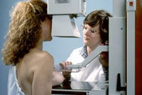
Mammography is the process of using low-dose X-rays to examine the human breast. It is used to look for different types of tumors and cysts. Mammography has been proven to reduce mortality from breast cancer. No other imaging technique has been shown to reduce risk, but self-breast examination (SBE) and physician examination are essential parts of regular breast care. In some countries routine (annual to five-yearly) mammography of older women is encouraged as a screening method to diagnose early breast cancer. Screening mammograms were first proven to save lives in research published by Sam Shapiro, Philip Strax and Louis Venet in 1966.
Like all x-rays, mammograms use doses of ionizing radiation to create this image. Radiologists then analyze the image for any abnormal growths. It is normal to use longer wavelength X-rays (typically Mo-K) than those used for radiography of bones.
At this time, mammography along with physical breast examination is still the modality of choice for screening for early breast cancer. It is the gold-standard which other imaging tests are compared with. CT has no real role in diagnosing breast cancer at the present. Ultrasound, Ductography, and Magnetic Resonance are adjuncts to mammography. Ultrasound is typically used for further evaluation of masses found on mammography or palpable masses not seen on mammograms. Ductograms are useful for evaluation of bloody nipple discharge when the mammogram is non-diagnostic. MRI can be useful for further evaluation of questionable findings, or sometimes for pre-surgical evaluation to look for additional lesions. Stereotactic breast biopsies are another common method for further evaluation of suspicious findings.
As Pravda.Ru previously reported Obese women with breast cancer are more likely to die from the disease than those of a normal weight, researchers said today.
US looked at information on more than 2,000 patients with early stage breast cancer between 1978 and 2003 who were treated with and radiotherapy.
They concluded that those who were classed as obese were more likely to die from breast cancer, despite it being diagnosed at an early stage.
They were also more likely to experience the spreading to another part of the body – metastatic disease, Scotsman reported.
According to the BBC News, the five-year survival rates for both normal weight and overweight patients was 92%, falling to 88% among those who were obese.
Lead researcher Penny Anderson said: "Our results show a statistically significant difference between obese women and the other groups.
"Because the prevalence of obesity increases with age, as does the risk of breast cancer, interventions that enhance weight control may have a substantial effect on breast cancer outcome."
Source: agencies
Subscribe to Pravda.Ru Telegram channel, Facebook, RSS!


